
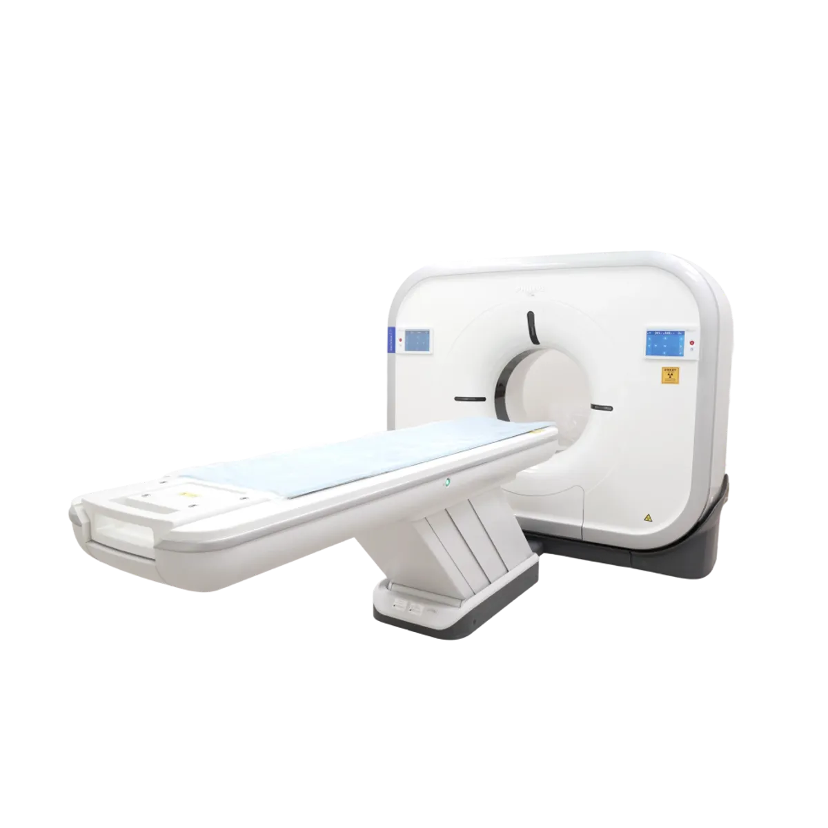
PHILIPS Incisive CT
PHILIPS It is an advanced computed tomography (CT) imaging system platform. This device combines X-ray technology with computer-based reconstruction techniques to produce high-quality cross-sectional images for medical imaging. CT provides cross-sectional views of the body’s internal anatomy in very thin slices and serves as a fundamental tool in the diagnosis of diseases.
PHILIPS Incisive CT What Kind of Technology?
Neurological and Head-and-Neck Imaging: Used to evaluate the brain, nervous system, and intracranial pathologies (e.g., stroke, tumor, hemorrhage). Philips Documents: Chest and Lung Imaging Used for evaluating lung nodules, infections, pulmonary embolism, and thoracic conditions. It can also be used for low-dose lung cancer screening protocols. Philips Documents: Cardiac Imaging Used for evaluating the heart and coronary arteries; specifically featuring software capabilities that reduce motion-induced artifacts.
Features
- Incisive CT captures multiple image slices using an X-ray tube and detectors that rotate within a gantry encircling the body, and uses a computer to generate 3D cross-sectional images.
- The device delivers clearer images using advanced algorithms and Philips’ AI-powered Smart Workflow technology, and helps optimize radiation dose.
- Philips’ “Tube for Life” warranty for Incisive CT may cover the free replacement of the X-ray tube under certain conditions for the duration of the system’s lifespan, thereby reducing operating costs.
- OnPlan patient-side gantry control: Allows technicians to control gantry movements from the patient’s side during the scan.
In Which Patients Is It Used?
Because it is a computed tomography (CT) system that caters to a wide range of clinical applications, it is used for diagnostic purposes in a wide variety of patient groups.